Thursday, 6 December 2012
Friday, 2 November 2012
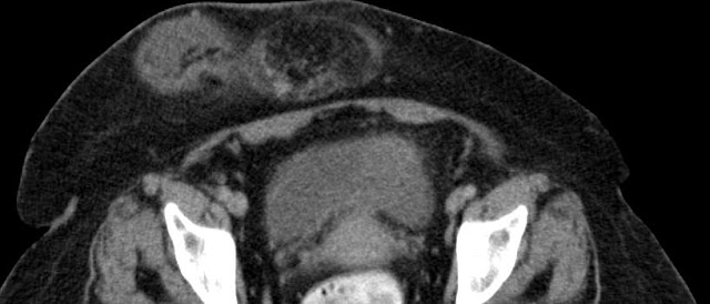
Left Ovarian Dermoid - Torsion
On USG - Right ovary was seen normally , Left ovary not visualized , right adenexal / iliac fossa complex mass lesion was seen.
On CT pedunculated heterogenously enhancing mass with soft tissue and fat densities - extending from left to right seen.
Surgically removed - biopsy report proved Ovarian Dermoid.
Case Followed up by : Dr. Peter Penuel Joshua , Dr. G.Chakradhar & DrAyushGoel
Saturday, 27 October 2012
Friday, 26 October 2012
Thursday, 25 October 2012
Wednesday, 24 October 2012
Tuesday, 23 October 2012
SDH
Acute on Chronic SubDural Hematoma
A subdural hematoma (American spelling) or subdural haematoma (British spelling), also known as a subdural haemorrhage(SDH), is a type of hematoma, a form of traumatic brain injury. Blood gathers within the outermost meningeal layer, between the dura mater, which adheres to the skull, and the arachnoid mater, which envelops the brain.
A subdural hematoma (American spelling) or subdural haematoma (British spelling), also known as a subdural haemorrhage(SDH), is a type of hematoma, a form of traumatic brain injury. Blood gathers within the outermost meningeal layer, between the dura mater, which adheres to the skull, and the arachnoid mater, which envelops the brain.
Subscribe to:
Comments (Atom)


